Fluorescence microscopy is based upon the concept that there are certain materials that emit energy which can be detected as a visible light. Each of these materials must be irradiated with a different specific light wavelength in order to cause an energy reaction in the form of light. The sample being used can either be treated with some fluorescing substances or it can be fluorescing on its original form.
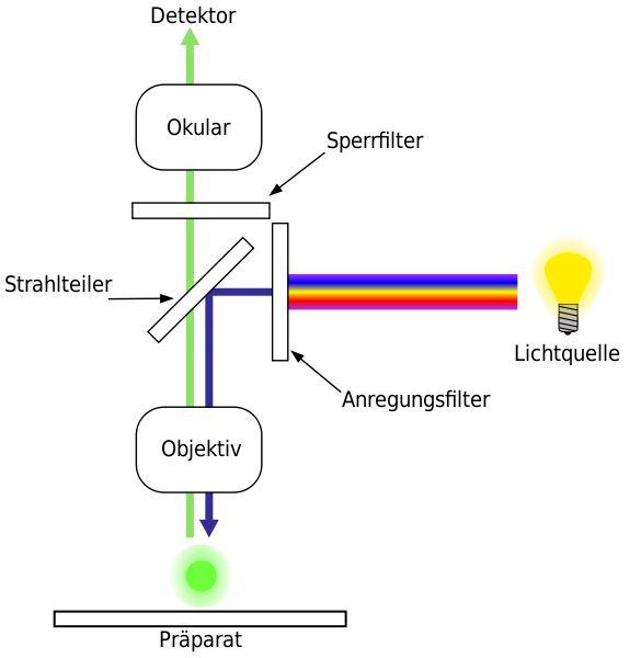
Fluorescence microscopy differs from most traditional techniques in that the visible light in the microscope eyepieces is not the original light emitted by the light source. The light you see is actually light that has fluoresced from the specimen itself. In order to receive such a response a high intensity light source must be used. This light is passed through a dichroic filter cube containing a fluorescence bandpass excitation filter, which only allows specific wavelengths of light to pass and reach the fluorescence specimen. After the incident filtered light reaches the specimen, it is no longer used, and any amount reflecting back into the microscope objective to the dichroic mirror is refined by the emission filter. The specimen fluoresces and it is this fluorescing light that passes back through the fluorescence emission filter and goes to the microscope eyepieces to provide a bright and colorful fluorescence image of the specimen. There are basically two types of illumination available in fluorescence microscopy, dark-ground condenser and epifluorescence. The dark-ground condenser simply facilitates the separation of the exciting light and the fluorescence light. With the introduction of epifluorescence though, it is now possible for the exciting light to reach the preparation level with the use of a dichroic mirror and condenser.
Epifluorescence microscopy (EFM) is a method of fluorescence microscopy that is widely used in life sciences. The basic task of an epifluorescence microscope is to get the sample excited. To do this, the excitatory light is passed from above through the objective lens and then onto the specimen instead of passing it first through the specimen. The fluorescence in the specimen gives rise to emitted light which is focused to the detector by the same objective that is used for the excitation. Since most of the excitatory light is transmitted through the specimen, only reflected excitatory light reaches the objective together with the emitted light and this method therefore gives an improved signal to noise ratio. An additional filter between the objective and the detector can filter out the remaining excitation light from fluorescent light. Because of the key features of an epifluorescence microscope, it is now becoming rapidly used in the biological and medical field. This microscopy technique has made it possible for people to determine and identify the cellular components of each sample with a high amount of specificity. For example, impurities and disease conditions can now be studied effectively by using epifluorescence microscopy. Also, many attachments are currently available for EFM's that are very helpful in research based practices. For example, the ApoTome system from Zeiss, Inc., (available at LMU) allows depth-discriminated images (optical slices) of fluorescence specimens to be produced. Compared to the conventional epifluorescence methods, these optical sections feature increased contrast and enhanced optical resolution in axial direction.