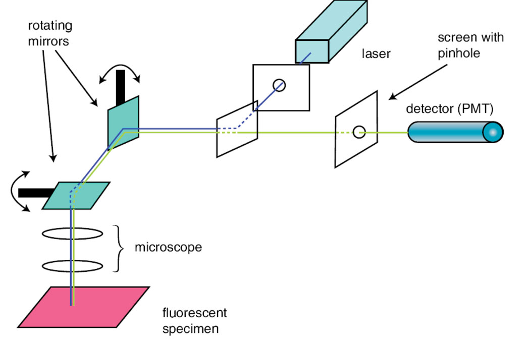
The Confocal Microscope (CM) is a simple yet considerably powerful variation of its predecessor, the conventional light microscope. In recent years the Confocal Laser Scanning Microscope has become widely established as a useful research instrument. The system procedure begins by exciting a specified point on its sample with a high-powered laser. The laser first passes through a series of mirrors which are used as filters and guides that scan the laser across the sample. Dye in the sample fluoresces due to the contact of the laser, and as a result the emitted light is de-scanned and focused into the pinhole. The emitted light is filtered and then measured by a computer-based detector. The computer collects every scanned point and manifests a two dimensional image.
At any given instant the microscope is only observing one point of your sample. Therefore, it is not a true image; it is simply the computer's compilation of several points rendered one pixel at a time. Focusing the microscope one point at a time may seem too drastic but this method coupled with a pinhole that is conjugate to the focal point (con-focal) makes this system extremely effective in rejecting out of focus fluorescent light, which is unavoidable in conventional scopes which illuminate the entire sample. The Confocal's abilities do not stop at 2-d renditions of a sample's surface however, it can also exclusively image a thin optical slice out of a thick specimen (optical sectioning), it can create a 3-dimensional image of the specimen using several optical slices as a 3D data set, and most impressively it can generate a time series of living specimens that undergo changes even in the range of microseconds.
In conclusion, the Confocal laser scanning microscopes are distinguished by their high spatial and temporal resolving power. They clearly outperform classical light microscopes especially by their axial resolution- a quality that enables users to acquire optical sections of a specimen.